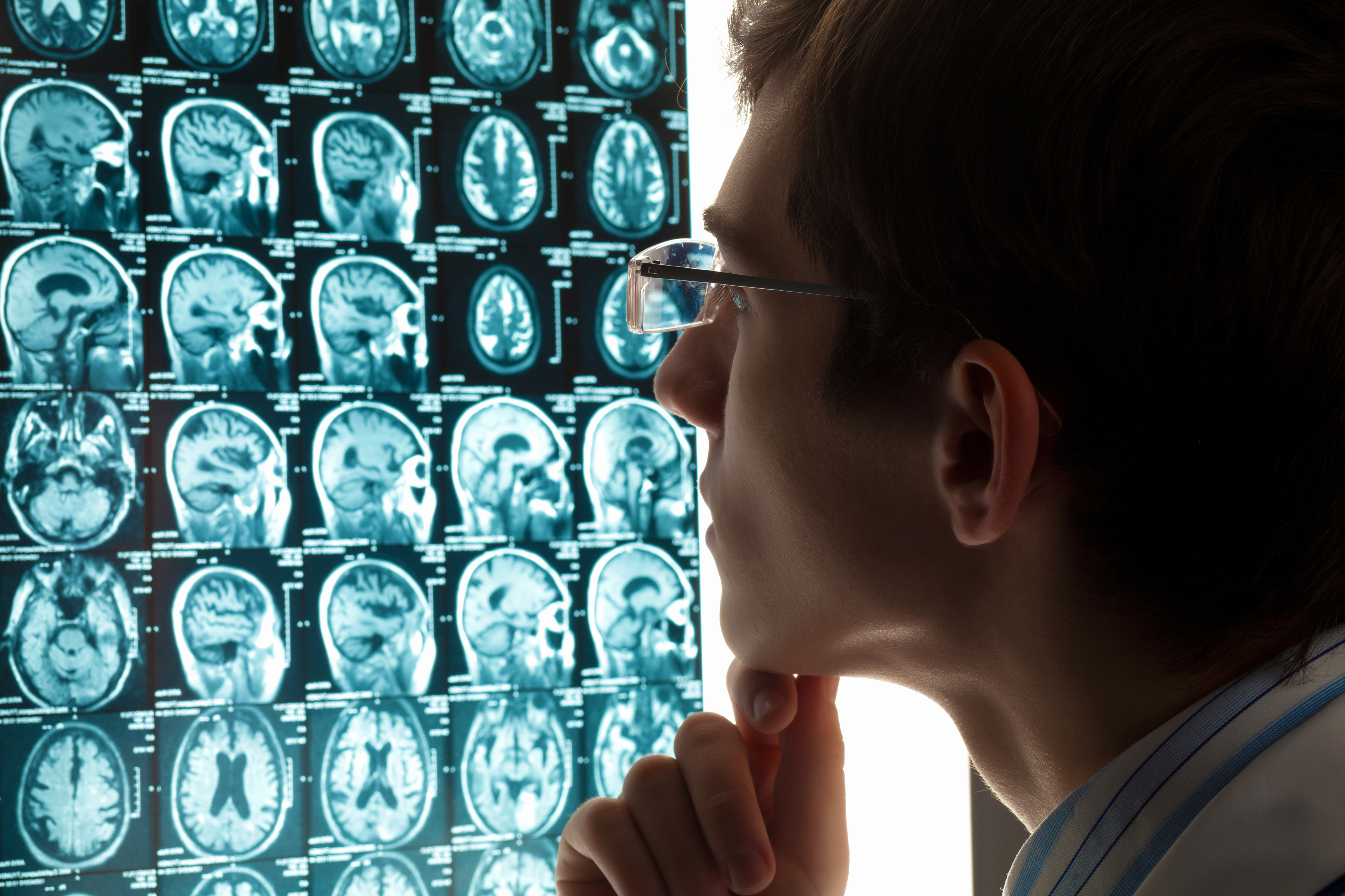
Brain MRI Can Detect Streaks of Blood Hours After Mild TBI

On March 20, 2013 neurologist Gunjan Parikh, M.D. presented a paper at the Annual Meeting of the American Academy of Neurology on MRI evidence of brain damage following mild TBI. Dr. Parkh works at the University of Maryland Shock Trauma Unit in Baltimore. He used MRI to evaluate 256 people with an average age of 50 who were admitted to the emergency department at Suburban Hospital in Bethesda and Washington Hospital Center in the District of Columbia after mild head injuries. Of those, 104 had imaging evidence of hemorrhage in the brain (67 percent reported loss of consciousness, and 65 percent reported amnesia, or temporary forgetfulness). Patients with hemorrhage underwent further MRI an average of 17 hours post-injury.

Two-third of these patients showed tiny, wide-spread micro-bleeds representing droplets of blood from broken brain capillaries. One-third showed streak-like or linear injuries which Dr. Parikh said represent a type of blood vessel injury similar to those seen in brains after severe head trauma. Dr. Parikh says that typically many patients with mild traumatic brain injury will not get this kind of scan. Accordingly MRI scans can be useful in finding patients who need additional treatment. Dr. Parikh has suggested making MRI standard protocol for evaluation of concussion patients.

Mild TBI Temporarily Raises Risk of Alcohol Addiction

A study published in the March 2013 issue of the American Journal of Psychiatry shows that mild TBI significantly increases the risk for alcohol addiction for a period of six months following injury with the risk gradually declining to normal thereafter. The findings come from a study of 5065 active-duty airmen who had sustained a mild TBI resulting in transient confusion or disorientation, memory loss, and/or brief loss of consciousness, and a comparison group of 44,733 airmen who had sustained other types of injury. After accounting for factors such as gender, marital status, ethnicity, age, deployment status, education level, rank, and career field, the team found that the risk for alcohol dependence was significantly increased in individuals with a mild TBI at 1-30, 31-179, and 180 days post diagnosis compared with those in the comparison group, at hazard ratios (HR) of 3.48, 2.66, and 1.70, respectively.

The study authors Whitehead et al. commented that: “Any alcohol use after TBI is concerning given the potential for reduction in spontaneous healing, risk of seizure or repeat TBI, and exacerbation of residual cognitive, emotional, and behavioral impairments.” They concluded that: “Screening for addiction-related disorders should be considered as part of routine care for mild TBI and might best capture the first 30 days post-mild TBI, with repeat alcohol screening thereafter for at least 6 months following the injury.” Families of people who have sustained a mild TBI should be vigilant in watching their drinking habits.

Pituitary Dysfunction after Pediatric TBI

According to an article in Volume 15, Issue 3 of the journal Pituitary published in Sept. 2012 there is evidence that about 30% of all children who suffer a traumatic brain injury show reduced pituitary output for up to 5 years post-injury. Hypopituitarism in children can be accompanied by reduced output of HGH (human growth hormone), stunted growth, and failure to develop normally during puberty. If the hypothalamus is damaged as a result of pediatric TBI the child can develop ACTH deficiency, diabetes insipidus, hypothyroidism or elevated prolactin. Given the serious consequences of endocrine disturbance post-TBI in children, it is recommended that children be monitored for such changes for at least one year post-TBI.

Allegation of Faking Depression in Litigation can be Disproven

Research published in October 2012 by a team at Mt. Sinai Medical Center led by Georgia Hodes Phd shows that real depression is related to and co-occurs with a measurable spike in the protein IL-6 produced by the immune system. In litigation over TBI claims the plaintiff is frequently depressed and the defense is either that the depression pre-existed the TBI (but was never diagnosed) or that the plaintiff is faking bad (i.e. pretending to be depressed to collect money he doesn’t deserve). Based on this published research if a plaintiff developed severe depression consequent to a TBI and his blood shows elevated IL-6 it will be easier to show that his depression is organic. In the rare case where the plaintiff had his blood tested shortly prior to the TBI and his IL-6 was measured as normal, you would have a strong pre vs. post incident contrast.

Zolpidem Wakes Some Patients From Persistent Coma

TBI and other conditions can put a person in a deep coma for years on end. This condition is called PVS which is short for “persistent vegetative state.” In a number of highly publicized incidents use of the drug Zolpidem (sold under the names Ambien, Ambien CR and Stilknox) has woken people up enabling them to sit up and speak after being in a PVS for years. Zolpidem is typically used to help people with insomnia get to sleep. It works by binding to the receptors for GABA, an inhibitory neurotransmitter. It has long been established that sleep and coma are different, and that coma is not a form of sleep. So how does Zolpidem wake people up from coma? We don’t know at this point. But the utterly remarkable success of Zolpidem in some cases has led people to call it the Lazarus drug.

Relationship of Sleep and Memory Disorders From TBI

Consequent to TBI some people suffer damage to an area of the brain called the Suprachiasmatic Nucleus or SCN. The SCN is about the size of a grain of rice and sits above the spot in the brain where the two sides of the optic nerve cross. It controls the 24 hour biological rhythym of sleep in humans and other mammals. Traumatic damage to the SCN disrupts normal sleep patterns and can also impair normal memory formation. This may be due to the fact that people need to sleep and dream a certain amount of time each night to form lasting memories. The function of the hippocampus (the brain organ of memory encoding) and the SCN are related. They share a certain number of genes in common. The matter is still under investigation.

Manage Pathological Laughing and Crying From TBI

One consquence of TBI known as the “psuedobulbar affect” or PBA is uncontrollable, inappropriate fits of laughter, crying or both. Jonathan Fellus, MD of Seacaucus, New Jersey, is an expert on the treatment of PBA. He recommends trying behavioral strategies first. For example, when you feel an episode coming you can try to relax, breathe, and distract yourself. If behavioral strategies do not work, then he recommends taking a medication called NUEDEXRA which can eliminate or decrease symptoms of PBA.

Human Growth Hormone Deficiency May Play a Role in Mild TBI Problems

Dr. Adriana G. Ioachimescu of Emory University, Atlanta presented a paper in September 2012 at the annual meeting of the Endocrine Society concerning the possible role of human growth hormone (HGH) deficiency in causing some problems for TBI suvivors. The subjects of her pilot study were 20 men (mean age, 34 years) with mild TBI resulting from military combat which had been sustained an average of 44 months earlier. Dr. Ioachimescu observed that all of the subjects had normal thyroid status and cortisol production, but 5 of them (25%) had a deficiency in their production of HGH.

While all 20 subjects with mild TBI performed similarly on measures of memory, learning, and simple and complex attention, the 5 subjects with deficient HGH did worse on tests of executive dysfunction, especially those relating to measures of inhibitory control and self-monitoring. The GH deficient soldiers show greater levels of depression and lower quality of life than the others even though they did not have a higher incidence of PTSD.

Was the deficiency of HGH present before the 5 veterans sustained their mild TBI or did it result from the MTBI? Either way the presence of deficient HGH in a person with MTBI appears to produce a worse outcome with regard to some executive functions and depressed mood. This suggests the possibility that treating MTBI survivors with poor executive function and/or depression using HGH may improve their condition. Futher research is needed.

Adult Stem Cell Therapy for TBI Still on the Horizon

In the Jan.-Feb 2009 issue of Disease Models & Mechanisms Dr. Peter A. Walker and colleagues summarized what we know so far about the use of progenitor cell therapies for traumatic brain injury. Due to evidence in rats that transplanation of embryonic stem cells grow tumors in the post-TBI brain and strict limits on the use of such cells, researchers have focused on adult stem cells. These cells congregate in certain areas of the adult human body, especially the bone marrow, the subventricular zone of the brain, the umbilicial cord and adipose (fat) tissue. There is preliminary evidence at this time that infusion of pluripotent adult stem cells into the brain post-TBI can protect damaged cells by supplying intact genetic material, producing neurotrophic growth factors, and by reducing inflammation.

Much more clinical experimentation needs to be done, but researchers are hopeful that one day adult stem cells will become part of the acute treatment for TBI. Right now there is little physicians can due in the acute stage except monitor intracranial pressure; reduce IC pressure with drugs, shunts or both; and increase cerebral blood perfusion with drugs.

One Cortisone Shot May Prevent PTSD

PTSD (post-traumatic stress disorder) can co-occur with TBI from the same event or occur on its own. Either way PTSD can cause serious emotional suffering and disability from work. A recent study on PTSD prevention by Joseph Zohar, M.D., of Tel Aviv University and Hagit Cohen, Ph.D., from Ben-Gurion University showed that traumatized patients who received a single injection of cortisone within a few hours of the event were more than 60% less likely to develop PTSD.

The researchers said the current prevention therapy using diazepine tranquilizers like Valium or Xanax to calm the traumatized patient down has not worked. They speculate the reason is that diazepines block the release of cortisol (which is made from cortisone). Why would blocking cortisol increase the likelihood of PTSD?

Cortisol increases blood sugar which reduces memory formation. Diabetics (who have high blood sugar) have poor cognitive function including poor memory. It may be that giving a shot of cortisone increases the material needed for cortisol synthesis which raises blood sugar and reduces memory of the event. Whatever the reason, the two researchers have been given a grant to continue studying the underlying mechanism(s).