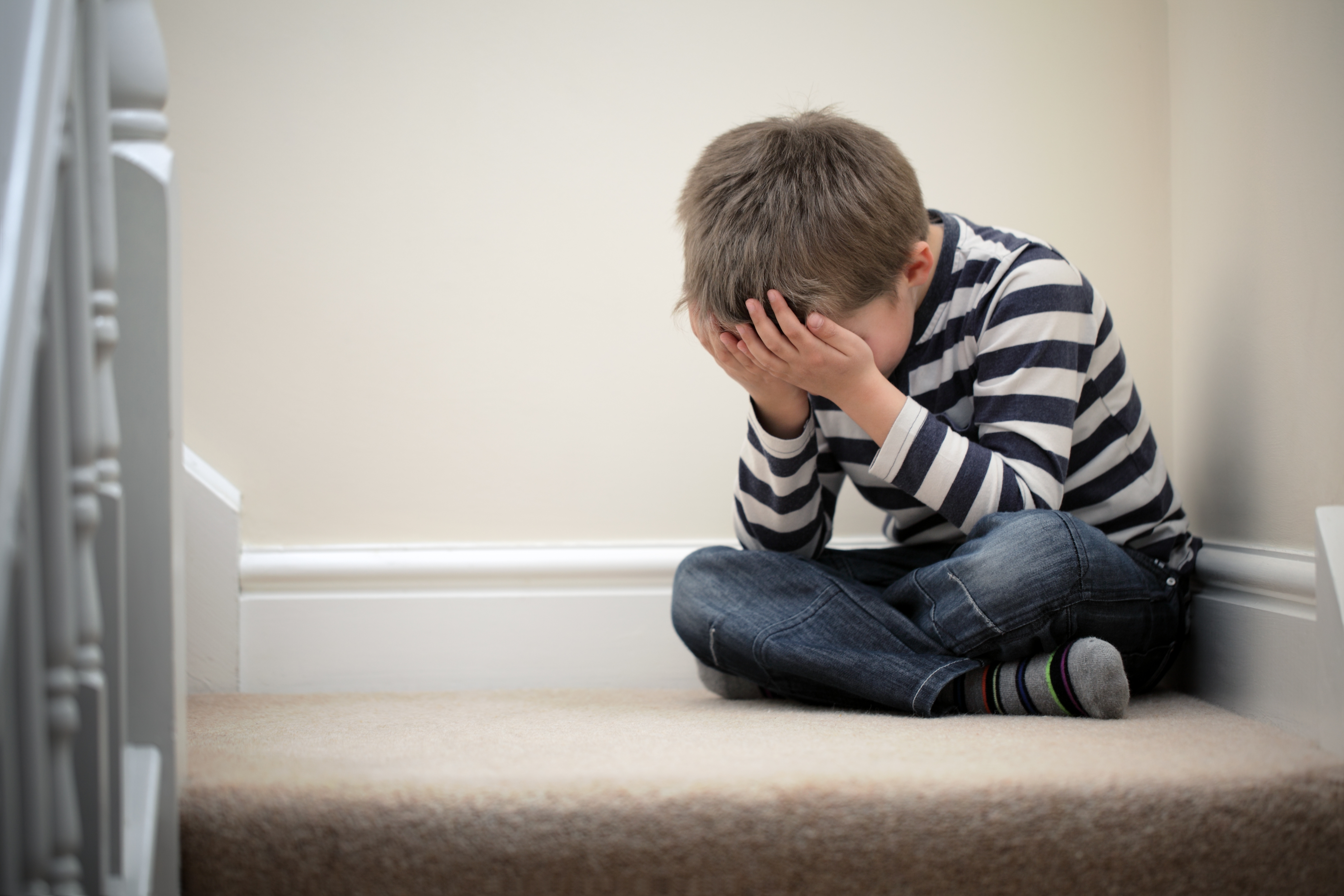
TBI Damages the Social Lives of Children Long After Injury

Neuropsychologist Shawn Gale, Ph.D. of Brigham Young University has just published a study of children three years after each one had suffered a frontal lobe TBI. He found that impairment of short term memory and cognitive processing speed made it very difficult for these children to carry on normal social conversation and social relationships. Although they looked fine these children ended up “loners” because they could not keep up with other children their age.

The study is available in the 4/10/14 issue of the Journal of Head Trauma Rehabilitation. Dr. Gale says it is very important to have children tested for these cognitive deficiencies and provided with cognitive therapy if they have them. A radiological correlate of this injury is a decrease in the thickness of the outer layer of the brain called the cerebral cortex.

TBI and Foreign Accent Syndrome

On January 14, 2014, a Canadian blog reported on the strange case of a Canadian mother of two named Sharon Campbell-Rayment who went from speaking with a Canadian accent to speaking with a Scottish accent following a TBI from falling off a horse. Sharon has been diagnosed by her doctors with foreign accent syndrome. This condition is extremely rare and only has 60 documented cases in the world. It is said to be only associated with traumatic head injuries or stroke and has sometimes been associated with people who have multiple sclerosis.

The syndrome is not completely understood. It may come from changes to timing, intonation and tongue placement after experiencing an injury. Another possibility is recovery of ancestral speech mechanisms. Sharon did research after developing her Scottish accent, and learned that her family had deep roots in Scotland and came to Canada one hundred years ago.

Coma Patients Appear to Recognize Faces of Loved Ones in New Study

On December 22, 2013 researchers working in Tel Aviv led by Dr. Haggai Sharon reported that patients in long term vegetative coma might be able to recognize familiar faces. Using functional magnetic resonance imaging (fMRI), they looked at brain activity in both healthy and vegetative subjects. First they showed both groups pictures of strangers, and then pictures of familiar faces. The brain activity patterns of the vegetative patients matched that of the healthy subjects.

According to Stanford neurologist Karen Hirsch all we know so far is that the facial recognition area of the brains of the coma patients was activated. Do they still know the people whose faces were shown to them and do they truly recognize those faces in a socially and emotionally meaningful way? Worst case scenario is that this is just a response to a stimulus with no other significance. At this point it is too early to tell, but research will continue. Dr. Sharon is hopeful that showing coma patients visual stimuli with emotional meaning may spark their brains and help push the healing process. This line of research, if successful, could potentially solve one of the most difficult problems of neurological critical care which is how do you know when someone is still living vs. brain dead? Sharon says” “If we could effectively communicate with the patient herself, perhaps we could simply ask whether or not she wants to go on living. However, we are still a long way from communicating with patients in such a way.”

Dog Saves Life of Vet with TBI and PTSD

War veteran Michael Sorenson who lives in the Chicago area was subjected to 4 separate blast injuries from IEDs in his service for the U.S. military in Iraq leaving him with TBI, PTSD, a spinal fracture, and severe depression. He received help in the form of Bane, a Pit Bull that was saved from euthanasia and retrained to be his therapy dog. One night Michael lay a knife on his bed but before he could use it to end his life Bane intervened and kept getting between him and the knife. When Michael realized Bane really cared about him he decided not to kill himself. Since then Michael and Bane have been inseparable. Michael even postponed back surgery to decrease his pain to pay for tendon surgery that Bane needed on his back leg.

The money to pay for that surgery came from good-hearted people and a good-hearted veterinarian who gave Michael a big discount. People living with TBI often fall into depression, sometimes severe enough to push them to contemplate suicide. Having a canine companion like Bane may be just the thing they need. Click on the link below to see Michael and Bane.

Scar Tissue From TBI Can Be Converted To New Functional Brain Cells

A research breakthrough in therapy to restore functional brain cells after TBI was just reported in the journal Cell Stem Cell on December 19, 2013. The discovery was announced by biologist Gong Cheng and his colleagues at Penn State University. Dr. Cheng was aware that after TBI glial cells rush to the area to clean up debris and protect vulnerable brain cells from infection. Unfortunately the glial cells form a scar which obstructs the growth of new neurons to replace the ones that died. Dr. Cheng likened it to fire, police, and ambulance personnel helping out the scene of an accident, but then getting stuck and blocking restoration of normal traffic. The ingenuous solution that Dr. Cheng’s team tested out whether they could get the glial scar tissue to convert to new brain cells by exposing them to a nerve growth factor, a protein called NeuroD1. The experiment involved causing TBI in adult mice, waiting for glial scar formation, and then using a retrovirus as a vehicle to safely carry NeuroD1 to the glial scar.

Remarkably just one week after inserting NeuroD1 into the glial scar tissue some of the glial cells (in particular the star-shaped astroglial cells and NG2 glial cells) were reprogrammed to convert into new brain cells. They then tried the same experiment on cultured human glial cells and got the same result. They found that the newly converted rodent neurons and human neurons were both capable of releasing and responding to neurotransmitters. Chen said “Our dream is to develop this in vivo conversion method into a useful therapy to treat people suffering from neural injury or neurological disorders.”

New Hope for TBI Survivors with Extreme Day Time Fatigue

A significant majority of people with TBI suffer from night-time insomnia and day-time fatigue. New research published in December 2013 by a group led by Akiva S. Cohen, Ph.D., a neuroscientist at The Children’s Hospital of Philadelphia (CHOP), may point the way to treatment. Dr. Cohen, in collaboration with other experts, induced TBI in mice which developed sleep disruption and day-time fatigue. The team fed these mice with selected branched chain amino acids (BCAA) which are precursors of the neurotransmitters glutamate and GABA. These neurotransmitters maintain a normal balance in brain excitation (wakefulness) and inhibition (sleep).

The researchers found that the new diet restored more normal patterns of sleep and wakefulness. On the BCAA diet the mice with TBI not only slept better at night but were much more active during the day. Their day-time EEG recordings showed marked improvement in activity level. Apparently the diet stimulated activity in the orexin neurons that keep the mice (and other mammals like humans) awake. After sustaining the TBI the mice showed decreased orexin levels in their spinal fluid, but these levels went up after eating the BCAA diet. Dr. Cohen cautioned Cohen cautioned that current evidence does not support TBI patients medicating themselves with commercially available amino acids from health food stores.

To help my readers understand the context of this research, the sleeping pill Ambien helps people get to sleep by activating the flow of GABA in the brain. Given the strange side effects reported by some Ambien users safety concerns exist regarding this drug even though it is currently the top seller in the sleeping pill market. The December 9, 2013 issue of the The New Yorker reported that Merck has developed a new sleeping drug called Suvorexant which works by inhibiting the orexin neurons which maintain wakefulness. This drug may be marketed in a year or so after further negotiations with the FDA. The BCAA diet Dr. Akiva’s team fed to mice activated the orexin neuron system in hopes of increasing day-time activity. TBI survivors with extreme day-time fatigue may wish to follow the progress of Dr. Cohen’s research.

DTI Scans Show Holes in Brain White Matter Years After Mild TBI

Mild TBI can occur to civilians from falls or car crashes and to military personnel from explosions which send shock waves through the brain. The microscopic damage to the brain that accompanies mild TBI is not visible on CT scans and standard MRIs. Because of this people with mild TBI are often dismissed as fakers, exaggerators or people with psychological problems rather than accepted as people with organic brain injury.

However, a special variety of MRI called DTI (water diffusion tensor imaging) can show microscopic brain damage by tracking leaks of water in the white matter tracts of the brain. Recently neurosurgeon Richard Bucholz, M.D. at St. Louis University Medical School became interested in what DTI would show in the brains of U.S. soldiers subjected to blast injuries years ago. On December 10, 2013 he reported to colleagues at the annual meeting of the Radiological Society of North America that DTI shows holes are still present in their brains from blast injuries. These soldier were still reporting problems with attention, memory, and mood. If you are still suffering from cognitive and/or mood problems years after a mild TBI and you have access to MRI using diffusion tensor imaging, you and your doctor may wish to consider using that type of scan, especially if you are being denied benefits for your injury.

Doctor examining a brain CT scan

New MRI Technique Shows Holes in Brain Membranes After Concussion

Up until now it has not been possible to visualize the effects of mild TBI on an MRI scan because standard MRI only picks up significant damage to brain tissue. However a new technique using MRI following injection of dye can demonstrate tiny holes in the meninges caused by a concussion, because the dye leaks through the holes. The meninges are the three thin membranes which snugly wrap the outside of the brain. Their job is to hold the brain in place, cushion the brain from minor shock, and keep out harmful molecules. The meninges contain an intricate set of blood vessels and nerves. It was established long ago that concussion can cause severe, post-traumatic headache by dilating blood vessels in the meninges and stretching the nerves that wrap around those vessels.

The new technique was developed by an ingenuous senior at Stanford University named Theo Roth who is majoring in biology. His work with humans (which was published online in the Journal Nature on December 8, 2013) derived from years of experimentation with mice in which Roth used a surgically implanted micro-camera to record the effects of concussion on mice brains in real time. Roth’s work was sponsored and made possible by Dorian McGavern’s lab at the National Institute of Neurological Disorders and Stroke (NINDS). Roth explains that when a concussion causes holes in the meninges it has damaged meningeal cells which leak internal substances (in particular something called reactive oxygen species) that are toxic to cells in the underlying brain tissue.

In the article in Nature Roth worked with co-scientist Lawrence Latour to examine 142 patients who have recently suffered a concussion but whose initial MRI scans had not revealed any physical damage to the brain tissue. Many of these patients were sent home from the hospital with the negative scans, but had since suffered headaches, memory loss or other hallmark symptoms of a mild brain injury. Latour injected the patients with a dye and conducted a follow-up MRI scan; in 49 percent of these patients, Latour saw the dye leaking through the meninges. This new technique is a true breakthrough for people suffering from mild TBI who are not believed by family, treating physicians or – in the case of litigation – by liability insurance companies and their defense lawyers.