PET Scans Show Brain Degeneration in Living NFL Football Players

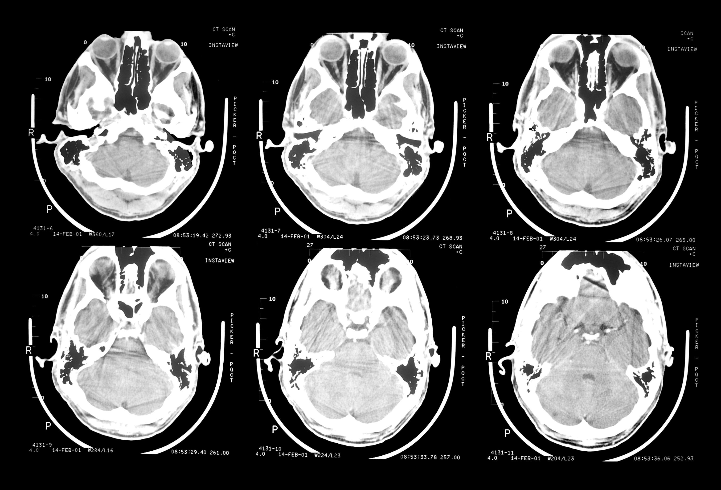
In the February 2013 issue of the American Journal of Geriatric Psychiatry, Dr. Gary Smalls and colleagues published research on their use of PET scans to search for abnormal deposits of tau protein in the brains of living people. Excessive tau has been correlated medically with such degenerative brain conditions as Alzheimer’s disease and CTE (chronic traumatic encephalopathy) seen in deceased NFL football players. CTE is associated with depression, memory loss, and violent outbursts. The brain of retired middle linebacker Junior Seau was proven to have CTE after it was donated to a neuropathologist by his family following Seau’s suicide.

In this study the researchers PET scanned 5 retired NFL players (aged 45-73) who had cognitive and mood problems and 5 age-matched controls. All NFL players showed significantly more accumulation of tau protein in their subcortical brain regions and amygdalas (the area of the brain involved in processing fear, anxiety, and anger). This study is the first time a scanning technique has been used to discern incipient degenerative brain disease in the brains of living athletes. It remains to be seen whether this technique can and will be applied to victims of TBI in other settings such as falls, car crashes and so forth.

TBI Rehab Centers Vary in Quality and Clinical Outcomes

Marie Dahdah, PhD, an investigator at the Baylor Institute for Rehabilitation in Texas, published a study 21 brain injury rehab centers in the United States in December 2013. Each of the centers was part of a national model systems program. At the outset of the study Dr. Dahdah expected to find fairly uniform results in clinical outcomes for patients with similar ages and injury severity. However there were significant variations, with a 25 percent to 45 percent difference between the best performing site and the site with the lowest outcomes at discharge. Dr. Dahdah plans to follow up with further investigation into which factors are responsible for these differences in outcomes so care can be improved across the country. The take home message for the family of a person needing TBI rehab is to do your homework rigorously on the facilities under consideration. Do not simply go on someone else’s word or settle for convenience.

New Use of Old Drug May Help Depression, Insomnia, and Fatigue Following TBI

Severe depression with insomnia and fatigue is relatively common following TBI. In patients with severe depression who were undergoing therapy and taking anti-depressants but showed only limited improvement, adding the wakefulness drug Modafinil produced a better outcome. This was especially true for the depressed patients in a group of 1,010 depressed patients for whom insomnia and fatigue were a major problem. This was the conclusion of researchers Barbara Sahakian from the University of Cambridge and Cynthia Fu from the University of East London. Their collaborative research study was published online in November 2013 in the Journal of Clinical Psychiatry.

Patients who do not benefit from antidepressants alone, and show significant sleep and fatigue problems, should consider exploring the benefits of adding Modafinil with their psychiatrist. Modafinil appears to have another advantage. Some patients with severe bipolar or unipolar depression are put on anti-psychotic medications that cause significant weight gain. In the April 1, 2009 issue of Biological Psychiatry Dr. James Roerig published a study showing that adding Modafinil reduced the gain of body mass index in patients taking an anti-psychotic drug.

Exposure to Just One Blast Shockwave Can Trigger Dementia Protein Formation

In the September 2013 issue of the Journal of Alzheimer’s Disease Dr. David Cook of VA Puget Sound Health Care System and University of Washington described some startling findings. Dr. Cook and his co-researcher Dr. Ibolja Cernak of the University of Alberta subjected mice to isolated shock waves similar to the kind suffered by American soldiers in Iraq and Afghanistan from IEDs. The magnitude of the shock waves was adjusted downward to reflect the much smaller size of the mice. What they found was that exposure to just one shock wave transformed tau protein in the mice brains to phospho-tau (the altered version of tau that tangles up the brains of symptomatic Alzheimer’s disease patients).

What is tau? Marc Kirschner at Princeton University discovered this molecule in 1975 and determined that its normal function is to stabilize the microtubules that provide structural integrity and some flexibility to the axons of brain cells as well as a means to transport nutrients from the axon tips to the nucleus of the cells. When the brain is subjected to a shockwave from a blow to the head or from exposure to a blast the tau protein molecules can fold in irregular patterns and form the tangles around axons associated with Alzheimer’s dementia. While it was known many years ago that repeated blows to the unprotected head of a boxer could cause dementia, and more recently that repeated blows to the helmet of an NFL football player, now we are seeing that just one exposure to a mild blast can cause tau tangles.

Beta-Amyloid is Deposited within Hours of Moderate to Severe TBI

Neuropathologists such as Bennet Omalu, M.D. and Ann McKee, M.D. have already established that NFL football players who had many concussions and who suffered from the cognitive, memory, personality, and behavioral changes consistent with CTE (chronic traumatic encephalopathy) had significant deposits of the beta-amyloid protein seen in Alzheimer’s disease when they died. What about people who have just one significant concussion? We now have a startling answer. On November 11, 2013 neuroscientist Hong Young published a study titled Amyloid Imaging With Carbon 11–Labeled Pittsburgh Compound B for Traumatic Brain Injury in JAMA Neurology online (doi:10.1001/jamaneurol.2013.4847). Dr. Young had PET scans performed on 15 persons (aged 21-50) with moderate to severe TBI and 11 healthy age-matched controls who had no signs, symptoms or history of neurologic disease. The results of the PET scans showed significant deposits of beta-amyloid protein in the cortical gray matter and striatrum (the brain’s motivation center) beginning within hours of the TBI in the TBI group, and an absence of such deposits in the brains of the healthy controls.

Although the size of the study was small and this is only study, it is important not just because it refutes the NFL position that even dozens of concussions do not cause brain changes associated with dementia, but because it alerts victims of even one significant concussion to their need for proper diagnosis and initiation of anti-amyloid therapy if significant amyloid deposits are found.

Decline in Social Status Post TBI Affects Brain Recovery

TBI is a form of brain damage that decreases the number of healthy brain cells and disrupts functional brain networks. If it is serious enough a TBI can cause disability with job and income loss leading to loss of socio-economic status and self-esteem. When you can’t work and you can’t pay your bills it is a sad fact of life that many people will treat you less well. It turns out that social stress actually decreases the ability of the brain to produce new cells to replace the ones that have died or suffered damages.

At the Annual Meeting of the American Academy of Neurology this month the Society for Neuroscience presented scientific evidence showing that people and animals under social stress (due to living in chaotic environments or being forced into submissive roles in the social hierarchy by more dominant beings) show brain abnormalities. The new findings include the following:

  • Adult rats living in disrupted environments produce fewer new brain cells than rats in stable societies, supporting theories that unstable conditions impair mental health and cognition
  • People who have many friends have certain brain regions that are bigger and better connected than those with fewer friends. It’s unknown whether their brains were predisposed to social engagement or whether larger social networks prompted brain development
  • Defeats heighten sensitivity to social hierarchies and may exacerbate brain activity related to social anxiety

“Social subordination and social instability have been associated with an increased incidence of mental illness in humans,” said press conference moderator Larry Young, PhD, of Emory University, an expert in brain functions involved with social behavior. “We now have a better picture of how these situations impact the brain.” What does this mean for TBI survivors? For those who have access to rehabilitation services that can return them to useful, personally meaningful work it makes sense to go for it, because returning to work may decrease anxiety and promote healthy brain repair. What about people who are too injured to return to work? It is possible to decrease anxiety through psychotherapy, meditation practice or prayer practice designed to increase self-acceptance. From experience I know that a person can find something valuable within herself even she cannot work even if this requires help from a psychologist, mental health counselor, clergy or chaplain.

traumatic brain injury in sports

Use Electricity to Treat Migraine Pain

The human brain contains mu-opioid receptors which block pain when activated by substances including mu-opioid and morphine. Alex DaSilva, assistant professor of prosthodontics at the University of Michigan has discovered that applying low dose electricity to the brain through electrodes on the scalp can trigger the release of mu-opioid and block certain types of head and facial pain, including migraine. Sessions take 20 minutes. To target the a safe, effective dose of electricity to the right place he uses a portable device called a high definition transcranial direct current stimulator. Migraine can be a big problem for people who suffer TBI. To find out more about DaSilva’s work you can follow him on the Internet through the pages of the Headache and Orofacial Pain Effort Laboratory at the U-M School of Dentistry.

TBI Quintuples the Risk of Depression in Normal Children

On October 25, 2013 Matthew C. Wylie, MD, presented a paper to the American Academy of Pediatrics called “Depression in Children Diagnosed with Brain Injury or Concussion.” Using data from the 2007 National Survey of Children’s Health, Dr. Wylie identified more than 2,000 children with brain injuries, reflecting the national child brain injury rate of 1.9 percent in 2007; and 3,112 children with diagnosed depression, mirroring the 3.7 percent national child depression rate that year. Compared to other children, 15 percent of those with brain injuries or concussions were diagnosed as depressed — a 4.9 fold increase in the odds of diagnosed depression. According to Dr. Wylie: “After adjustment for known predictors of depression in children like family structure, developmental delay and poor physical health, depression remained two times more likely in children with brain injury or concussion.”

Using PET Scans to Diagnose Mild TBI

CT scans and MRIs do not show mild TBI because mild TBI exists at the microscopic level unlike severe or moderate TBI which are accompanied by macroscopic bruising, swelling or bleeding of brain tissue. Some clever neuroscientists at the University of Virginia have just found a way to detect mild TBI using a PET scanner. The technique involves putting a radioactive tracer in the patient’s blood which attaches itself to neutrophils (white blood cells that travel through blood vessels which access cerebro-spinal fluid to the site of a brain injury). When a PET scan is taken of the brain of a patient with a mild TBI the neuro-radiologist ought to be able to see one or more areas where the radioactively traced neutrophils have gathered to clear the microscopic debris from the brain injury. This research was funded by the U.S. military’s Defense Health Program. The UVA research team consists of radiologist/neuroscientist James Stone, MD, PhD, and radiology researchers Stuart Berr, PhD, Jiang He, PhD, and Dongfeng Pan, PhD.

Research on TBI in Fruit Flies will Yield Data Applicable to Humans

University of Wisconsin geneticist Barry Ganetzky concluded that we know very little about how head trauma triggers neurodegeneration of the brain following TBI, and that’s because of legal and ethical restrictions on human experimentation. After looking around for a suitable non-human candidate for TBI research he hit on the fruit fly. The fruit fly’s brain is encased in a hard cuticle which mimics the human skull, and when trauma is applied to its cuticle the fruit fly not only suffers short term effects of concussion, but delayed onset of neurodegenerative changes to its brain just like some human victims of TBI.Ganetzky and his colleague David Wassarman, a UW professor of cell and regenerative biology, have received an NIH grant to pursue their research. Their first report came in the October 14, 2013 issue of the Proceedings of the National Academy of Sciences. As they continue to report on their work I will relay it on this blog.